Understanding all about the X-Ray Machines
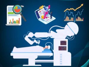
X-ray machines are amazing tools that have transformed medicine and research in amazing ways. They are giving us precise insight into the insides of the human body and are changing the way we diagnose and treat various diseases. X-ray images account for more than 60% of all diagnostic imaging worldwide. This highlights the importance of X-ray images in medicine.
Beyond diagnosis, X-ray machines also visualize internal structures, guide interventions, and aid fields such as material science and archaeology. They have greatly improved patient care, advanced medical research to unprecedented levels. Eventually demonstrated their essential role in medical and scientific research.
Show how X-ray machines revolutionized medical and scientific research
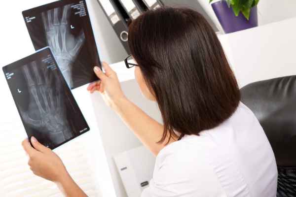
The discovery of X-rays by Wilhelm Conrad Roentgen in 1895 revolutionized research and medicine. A chance experience with cathode rays led Roentgen to observe these mysterious rays, which allowed him to obtain the first X-ray images of his wife’s hand bones. His breakthrough transformed medicine by allowing noninvasive visualization of internal structures to diagnose fractures, tumors, and abnormalities. Advances in X-ray technology have improved image clarity and accuracy over time.
The X-ray instrument has played an important role in non-diagnostic research, contributing to the understanding of DNA structure and atomic interactions. Its range of applications includes dentistry, veterinary medicine and security, reinforcing its significant influence in both fields.
What are the differences between early and modern X-ray machines?

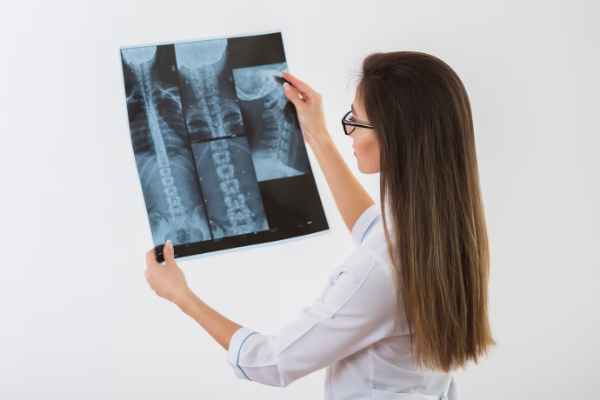
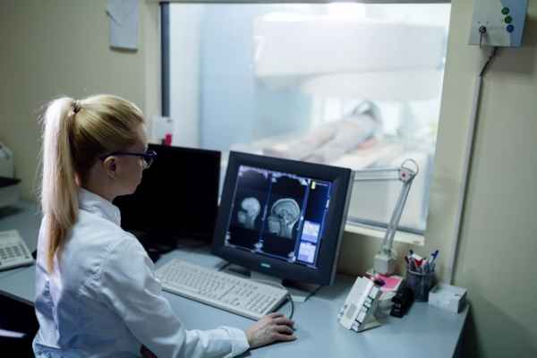
X-ray equipment has evolved significantly from early models to modern digital imaging systems, transforming clinical diagnosis. Early machines developed in the late 19th and early 20th centuries had limited performance and low image resolution. By the mid-20th century, fluoroscopy enabled real-time imaging and improved dynamic visualization. An important milestone was the introduction of computed tomography (CT) in the 1970s, which provides cross-sectional images of the body. Digital radiography and mammography replaces film-based systems, offering faster image acquisition, better quality, and electronic storage. These advances have revolutionized medical imaging, allowing for accurate diagnosis and improved patient care.
Applications of X-ray machines in medicine
-
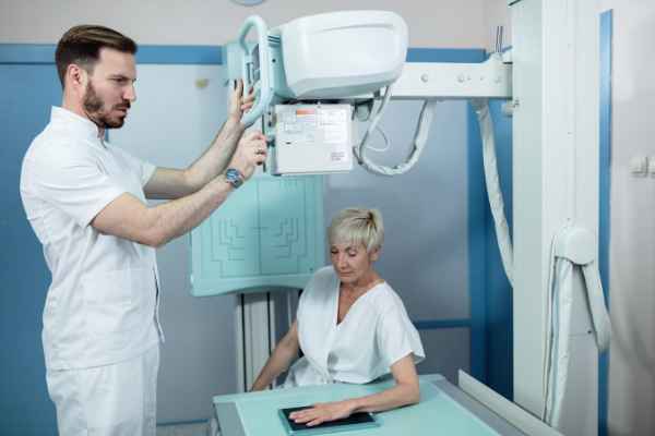
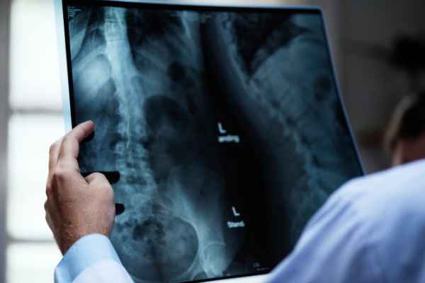
- Diagnostics: X-ray equipment is essential for diagnostic imaging, detecting fractures, tumors, infections, and other abnormalities, assisting in treatment decisions, and monitoring disease progression.
- Interventional Procedures: X-ray equipment guides minimally invasive procedures such as angiography and fluoroscopy, and provides real-time imaging during catheter placement and joint injections.
- Dental: X-ray equipment is commonly used in dental offices to examine tooth and jaw structure, identify cavities, determine tooth position, and assist in treatment planning.
- Radiotherapy: In radiation therapy, an X-ray machine uses a linear accelerator to deliver a targeted dose of radiation to a cancerous tumor, minimizing damage to surrounding healthy tissue
- Research and Education: X-ray equipment is an essential tool in medical research, allowing the study of the structure and function of various biological systems to improve anatomy, physiology, pathology, and molecular biology.
X-ray machine contributions to research
-
- Structural Biology: X-ray crystallography, using X-ray diffraction patterns, reveals the atomic and molecular structure of biological macromolecules, such as proteins and DNA, and helps us understand their functions and interactions.
- Materials Science: X-ray equipment analyzes material structure and determines crystal structure for the development of new materials with specific properties important for electronics, energy storage, and engineering.
- Archaeology and Paleontology: X-ray imaging revolutionizes the non-destructive study of ancient artifacts and fossils, allowing for the precise analysis, preservation and understanding of historical and paleontological items Helpful.
- Medical Imaging Research: X-ray equipment helps improve the quality of medical images, reduce radiation doses, and develop new modalities for more accurate body imaging.
- Fundamental Physics Research: X-ray equipment is essential for physics research, including particle physics and astrophysics. They use synchrotron radiation facilities to study the properties of matter and investigate particle interactions.
Innovations in X-ray technology and their future possibilities
-
- Digital Imaging: The transition from film-based to digital X-ray systems has revolutionized the capture, storage, and analysis of images, increasing quality while reducing radiation dose.
- 3D Imaging: Advancements such as cone beam computed tomography (CBCT) have enabled 3D imaging, improving diagnostic capabilities in dentistry, orthopedics, and interventional radiology.
- Artificial Intelligence (AI) Integration: AI algorithms integrated into X-ray equipment support faster and more accurate diagnosis by interpreting images, detecting abnormalities, and optimizing image parameters.
- Spectral Imaging: Techniques such as dual energy imaging enable further characterization of tissues, improving structural differentiation and pathology detection.
- Reducing Radiation Dose: Ongoing research is focused on minimizing radiation dose while maintaining image quality through optimized protocols and advanced reconstruction techniques.
- Portable and Point-of-Care X-ray: Advances in miniaturization and wireless technology enable portable X-ray systems that facilitate imaging in remote environments and enhance point-of-care applications.
- Fusion Imaging: Integrating X-ray with other modalities such as ultrasound and MRI improves diagnostic accuracy and supports minimally invasive procedures by providing complementary information.
Sum up,
Advances in X-ray technology promise exciting possibilities for the future. These include digital imaging, 3D capabilities, AI integration, spectral imaging, radiation dose reduction, wearable systems, and fusion imaging. These improvements aim to increase diagnostic accuracy, improve patient care, and expand applications across various medical specialties.